Scientific facts about llamas: elliptical blood cells, nanobodies and physiology
Anatomy and physiology of Lama glama: elliptical red blood cells adapted to Andean hypoxia, heavy-chain antibodies (VHH nanobodies), three-chambered stomach and biomechanics.
TodoLlamas · Editorial approach
Published · Content rewritten
Beyond its historical importance as an Andean pack animal and fibre producer, the llama (Lama glama) is a remarkable model organism for comparative physiology, molecular immunology and veterinary science. Its evolutionary lineage combined tens of millions of years of adaptation to arid plains with a subsequent colonisation of the high Andean puna—an environment often situated between 3,700 and 5,000 metres above sea level where atmospheric oxygen partial pressure drops to roughly half its sea-level value, ultraviolet radiation is intense and vegetation consists of tough, seasonal grasses. [1, 5] To thrive under these pressures, the llama evolved haematological, immunological, digestive and biomechanical adaptations unique among placental mammals.
Elliptical red blood cells and high-affinity haemoglobin
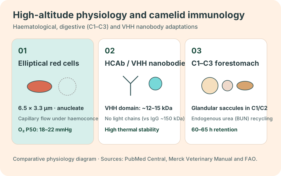
In almost all mammals, mature erythrocytes (red blood cells) are circular, biconcave discs. Camelids represent the major exception within the class Mammalia: their mature erythrocytes are small, flat, non-nucleated and distinctly elliptical or oval in outline (measuring roughly 6.5 × 3.3 micrometres). Maintained by a specialised spectrin cytoskeleton and marginal microtubular band, this elliptical geometry confers two vital physiological advantages. First, flat oval cells align smoothly with laminar blood flow and traverse narrow capillary beds without sludging when dehydration increases plasma viscosity. Second, they exhibit extraordinary osmotic resistance, allowing a thirsty camelid to drink large volumes of water rapidly without triggering osmotic haemolysis (rupture) of its red blood cells.
This cellular morphology works in tandem with a biochemical adaptation in the haemoglobin molecule. When lowland mammals ascend to high altitude, they compensate for hypoxia by overproducing red blood cells (secondary polycythaemia), which thickens the blood, raises pulmonary arterial pressure and strains the right heart ventricle. By contrast, llamas maintain a modest haematocrit (around 27% to 35%) even above 4,000 metres. Their haemoglobin possesses an exceptionally high intrinsic affinity for oxygen—reflected in a left-shifted oxygen dissociation curve and a low P50 value—enabling efficient oxygen loading in thin mountain air without pathological blood thickening. [1, 4]
Heavy-chain antibodies and nanobodies (VHH) in biotechnology
One of the most influential discoveries in modern molecular immunology emerged from studying camelid blood serum. In humans and most other vertebrates, conventional immunoglobulin G (IgG) antibodies are heterotetrameric proteins assembled from two heavy chains and two light chains (~150 kilodaltons). Alongside conventional IgGs, however, the immune system of llamas, alpacas and Old World camels produces fully functional heavy-chain-only antibodies (HCAbs) that completely lack light chains as well as the first constant domain (CH1).
Each heavy chain in these special antibodies binds antigens through a single variable domain known as VHH or a nanobody. With a molecular mass of only 12 to 15 kilodaltons—roughly one-tenth the size of a conventional antibody—the llama VHH fragment is the smallest naturally occurring intact antigen-binding unit known to science. Because its complementarity-determining loops (particularly the extended CDR3 loop) project outward like a finger rather than forming a flat surface, a nanobody can reach deep into hidden viral pockets, enzyme active sites and cryptic receptor clefts that standard antibodies cannot access. Llama nanobodies also display remarkable thermal and chemical stability, resist extreme pH levels and can be produced economically in microbial cultures for antiviral therapeutics, cancer imaging and antivenom research.
Pseudoruminant digestive physiology and urea recycling
Although llamas regurgitate and rechew their cud and ferment plant cellulose, they do not belong to the taxonomic suborder Ruminantia (cattle, sheep, goats and deer); instead, they belong to the suborder Tylopoda. [1, 4] This convergent evolution is evident in their stomach anatomy, which consists of three interconnected compartments labelled C1, C2 and C3 rather than the four chambers (rumen, reticulum, omasum and abomasum) of true ruminants. [3]
The first compartment (C1) accounts for over 80% of gastric volume and acts together with the smaller second compartment (C2) as an anaerobic fermentation vat. Unlike the keratinised papillated lining of a cow's rumen, the ventral walls of C1 and C2 contain specialised glandular saccules (everted mucosal pouches) whose bicarbonate-rich secretions buffer volatile fatty acids generated during microbial fermentation. The third compartment (C3) is an elongated tubular organ whose distal fifth houses true gastric glands that secrete hydrochloric acid and pepsin. [2, 3]
By combining vigorous cyclical forestomach contractions with a long mean retention time for fibrous particles (often 50 to 60 hours, compared with 30 to 40 hours in sheep), llamas extract more digestible energy from coarse, lignified highland grasses. They also possess a highly efficient nitrogen-conservation pathway: when dietary protein drops during the dry season, the kidneys sharply curtail urinary urea excretion and return plasma urea into C1 via saliva and directly across the forestomach wall, where symbiotic microbes convert it into high-value microbial protein. See our guide on what llamas eat for how this digestive physiology informs forage and mineral management.
Specialised dentition, split upper lip and fighting teeth
A llama's oral anatomy is tailored for selective browsing and grazing without uprooting fragile pasture plants. Its upper lip is divided by a deep median cleft (philtrum) into two independently mobile, prehensile halves capable of sorting tender leaves from thorny stems or stony ground. [1, 2] In the lower jaw, six procumbent spatulate incisors shear vegetation against a tough, fibrous upper dental pad rather than upper central incisors.
Adult camelids retain a striking dental structure absent in cattle and sheep: sexually dimorphic “fighting teeth”. [2] Consisting of six sharp, backward-curved caniniform teeth—one modified upper third incisor (I3), one upper canine and one lower canine on each side of the jaw—these weapons erupt fully in intact males between two and three years of age. Males use them during dominance battles to slash at a rival's legs, neck and scrotum, an evolutionary trait traced in our article on camelid evolution and history.
Plantar foot pads, pacing gait and the thermal window
| Biological system | Anatomical or molecular trait | Primary adaptive function |
|---|---|---|
| Haematological and respiratory | Flat, non-nucleated elliptical erythrocytes and high-affinity haemoglobin. | Efficient oxygen transport at 3,500–5,000 m without hyperviscosity, plus high osmotic resilience. |
| Immunological | Heavy-chain-only antibodies lacking light chains (VHH nanobody domains). | Binding of recessed or cryptic antigenic epitopes with exceptional thermal and chemical stability. |
| Digestive (Tylopoda) | Three-chambered stomach (C1, C2, C3) with glandular saccules and urea recycling. | Superior fermentation of low-protein fibrous forage and continuous intragastric buffering. |
| Locomotor and pedal | Two-toed foot with leathery digital pads (tylium), toenails and pacing gait. | Sure-footed grip on rocky trails, reduced soil compaction and metabolic energy conservation. |
| Thermoregulatory | Double-layered dorsal fleece paired with a sparsely haired ventral thermal window. | Insulation against sub-zero nights combined with posture-controlled heat dissipation. |
The suborder name Tylopoda literally means “padded foot”. Instead of standing on the tip of the third phalanx encased in a rigid keratin hoof wall (unguligrade stance), a llama supports the middle and distal phalanges of its two digits (III and IV) on resilient fibro-elastic digital cushions covered by a leathery plantar sole, with small curved toenails at the front tips. [1, 5] When moving at speed, llamas employ an ipsilateral pacing gait, advancing both legs on the same side of the body simultaneously to lengthen stride and minimise vertical oscillation of the pack load.
Finally, llamas cope with daily Andean temperature swings exceeding 25 °C by pairing a thick insulating dorsal coat with a “thermal window” across the ventral abdomen, armpits and inner thighs, where skin is sparsely haired and richly supplied with superficial blood vessels and sweat glands. [2] On freezing nights, the llama folds its legs beneath its body in sternal recumbency (kush posture), sealing the thermal window against the ground; during hot afternoons or in warm lowland climates, it stands or extends its limbs in the shade to radiate and convect excess body heat. Compare how coat structure and body frame vary across all four Andean species in our identification guide to llamas, alpacas, vicuñas and guanacos.
Frequently asked questions about llama biology
- Do llamas have four stomachs like cows? No; llamas are pseudoruminants with a single stomach divided into three interconnected compartments (C1, C2 and C3) lined in part with glandular saccules. [2, 3]
- Do llama red blood cells have a nucleus? Mature circulating llama red blood cells are non-nucleated, just like those of other mammals, but they are uniquely flat and elliptical rather than round and biconcave.
- Why are llama nanobodies important in human medicine? VHH nanobodies derived from camelid heavy-chain antibodies are used in biomedical research and clinical pharmacology to neutralise respiratory viruses, block toxins, stabilise membrane proteins for structural imaging and target cancer cells.
Sources and references
- University of Michigan, Animal Diversity Web: Lama glama
- Merck Veterinary Manual: Management of Llamas and Alpacas
- Merck Veterinary Manual: Stomach, llama
- Fan et al. (2020): Genomic analysis of the domestication and post-Spanish conquest evolution of the llama and alpaca
- FAO: Alpaca and llama husbandry manual (Spanish), introduction and distribution